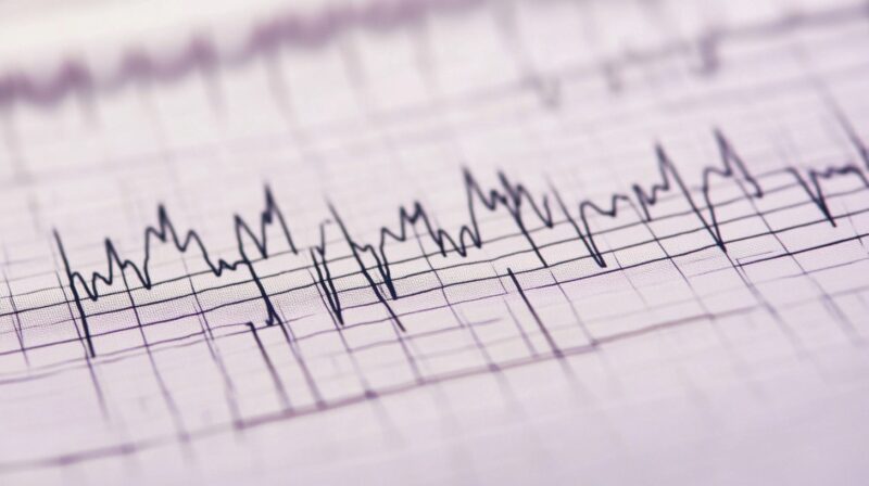
Being aware of ECG/EKG rhythms is vital for nurses, especially when preparing for the NCLEX exam.
It is just one of many things nurses need to know about before they can conduct their job properly.
There are key arrhythmias and heart conditions that nurses must recognize quickly to ensure prompt and appropriate treatment.
With that in mind, we wanted to provide you with 10 essential heart rhythms and their characteristics that nurses find particularly useful in their line of work.
1. Ventricular Fibrillation (V-Fib)
Ventricular Fibrillation (V-Fib) is a life-threatening arrhythmia where the ventricles quiver ineffectively, causing the heart to stop pumping blood. It is a medical emergency that often leads to sudden cardiac arrest.
- Cause: Often a result of myocardial infarction, electrical shock, or severe electrolyte imbalances.
- ECG Characteristics: There are no discernible P waves or QRS complexes; the waveform appears chaotic and irregular.
- Symptoms: Loss of consciousness, no pulse, no blood pressure, and cessation of breathing.
- Treatment: Immediate defibrillation is crucial to restore a regular rhythm. Cardiopulmonary resuscitation (CPR) should be performed while preparing the defibrillator. Medications such as epinephrine and antiarrhythmics like amiodarone may be administered.
V-Fib can lead to death within minutes if untreated. Recognizing the ECG pattern promptly is critical for effective intervention.

2. Ventricular Tachycardia (V-Tach)
Ventricular Tachycardia (V-Tach) is a rapid heart rate originating from the ventricles, often more than 100 beats per minute. It can be stable (with a pulse) or unstable (without a pulse).
- Cause: Commonly associated with heart disease, electrolyte imbalances, or ischemia.
- ECG Characteristics: Wide and regular QRS complexes, with no discernible P waves. The rate is usually between 150-250 beats per minute.
- Symptoms: Dizziness, palpitations, shortness of breath, or unconsciousness if the patient is unstable.
- Treatment: For stable V-Tach, antiarrhythmic medications like amiodarone or procainamide may be given. In unstable V-Tach, immediate cardioversion or defibrillation is necessary.
Early recognition of V-Tach can prevent progression to more dangerous rhythms like V-Fib.
3. Torsades de Pointes

Torsades de Pointes is a specific type of ventricular tachycardia characterized by twisting of the QRS complexes around the isoelectric line. It is often associated with prolonged QT intervals.
- Cause: Prolonged QT interval due to medications, electrolyte imbalances (especially low magnesium), or congenital conditions.
- ECG Characteristics: Polymorphic QRS complexes that appear to twist around the baseline. The rhythm is irregular.
- Symptoms: Palpitations, dizziness, syncope, and in severe cases, sudden cardiac arrest.
- Treatment: Intravenous magnesium sulfate is the treatment of choice. Cardioversion may be needed if the patient is unstable. Removing the causative agent (e.g., certain medications) is also important.
Torsades are often transient but can progress to V-Fib if not treated promptly.
4. Supraventricular Tachycardia (SVT)
Supraventricular Tachycardia (SVT) is a rapid heart rate originating above the ventricles, often exceeding 150 beats per minute. It includes rhythms such as atrial tachycardia and AV nodal reentrant tachycardia.
- Cause: Can occur due to stress, excessive caffeine, alcohol, or heart conditions like Wolff-Parkinson-White syndrome.
- ECG Characteristics: Regular, narrow QRS complexes, with P waves often hidden in the preceding T waves.
- Symptoms: Palpitations, shortness of breath, anxiety, chest pain, and lightheadedness.
- Treatment: Vagal maneuvers (e.g., carotid massage) are the first-line treatment. If ineffective, adenosine is given to slow the heart rate. Electrical cardioversion is used in unstable cases.
SVT can be uncomfortable but is usually not life-threatening if treated promptly.
5. ST-Elevation Myocardial Infarction (STEMI)

ST-Elevation Myocardial Infarction (STEMI) is a severe type of heart attack where there is complete blockage of a coronary artery, leading to significant heart muscle damage.
- Cause: Atherosclerosis leading to a complete coronary artery blockage.
- ECG Characteristics: ST-segment elevation in two or more contiguous leads, representing myocardial injury. May also show pathological Q waves and inverted T waves over time.
- Symptoms: Crushing chest pain, shortness of breath, diaphoresis, nausea, and radiating pain to:
- Arm
- Neck
- Jaw
- Treatment: Immediate reperfusion therapy is required, either through percutaneous coronary intervention (PCI) or thrombolytics. Aspirin, nitroglycerin, and anticoagulants are also commonly used.
Quick identification and treatment of STEMI is crucial to prevent permanent heart damage.
6. Atrial Fibrillation (A-Fib)
Atrial Fibrillation (A-Fib) is an irregular and often rapid heart rhythm originating from the atria. It can lead to poor blood flow and an increased risk of stroke.
- Cause: Hypertension, heart failure, valvular heart disease, or hyperthyroidism.
- ECG Characteristics: Irregularly irregular rhythm with no discernible P waves. The QRS complexes are narrow and irregular.
- Symptoms: Palpitations, fatigue, shortness of breath, and dizziness. Some patients may be asymptomatic.
- Treatment: Rate control with beta-blockers or calcium channel blockers, rhythm control with antiarrhythmics, and anticoagulation therapy to prevent stroke.
Chronic A-Fib requires long-term management to prevent complications like thromboembolism.
7. Premature Ventricular Contractions (PVCs)

Premature Ventricular Contractions (PVCs) are extra beats originating from the ventricles, disrupting the normal heart rhythm.
- Cause: Stress, caffeine, electrolyte imbalances, or underlying heart disease.
- ECG Characteristics: Early, wide, and abnormal QRS complexes, often followed by a compensatory pause.
- Symptoms: May feel like a skipped heartbeat or fluttering in the chest. Frequent PVCs can cause dizziness or fatigue.
- Treatment: Infrequent PVCs generally don’t require treatment. Lifestyle modifications, such as reducing caffeine, can help. Antiarrhythmic medications are used if PVCs are frequent and symptomatic.
While usually benign, frequent PVCs warrant further investigation.
8. Sinus Bradycardia (Sinus Brady)
Sinus Bradycardia is a slow but regular heart rate of less than 60 beats per minute originating from the sinus node.
- Cause: Can be a normal variant in athletes, or caused by medications (e.g., beta-blockers), hypothermia, or increased vagal tone.
- ECG Characteristics: Regular rhythm with normal P waves preceding each QRS complex, but at a slower rate.
- Symptoms: Fatigue, dizziness, fainting, or syncope. In some cases, it can be asymptomatic.
- Treatment: If symptomatic, treatment may include atropine or pacing. Stopping the offending medication can also help.
It may not always require intervention unless the patient is symptomatic.

9. Sinus Tachycardia (Sinus Tach)
Sinus Tachycardia is a regular but rapid heart rate of more than 100 beats per minute, originating from the same source as sinus bradycardia.
- Cause: Fever, stress, dehydration, anemia, or hyperthyroidism.
- ECG Characteristics: Regular rhythm with normal P waves preceding each QRS complex, but at an increased rate.
- Symptoms: Palpitations, dizziness, shortness of breath, and chest discomfort.
- Treatment: Treat the underlying cause (e.g., fever, dehydration). Beta-blockers may be used if the patient remains symptomatic after addressing the cause.
It is an absolute must to know that it is often a compensatory mechanism rather than a primary problem.
10. Normal Sinus Rhythm (NSR)

Normal Sinus Rhythm (NSR) is the ideal heart rhythm, with electrical activity originating from the sinus node.
- ECG Characteristics: Regular rhythm, rate between 60-100 beats per minute, with normal P waves before each QRS complex.
- Symptoms: No symptoms, as this is the normal functioning rhythm of the heart.
- Treatment: None required, as this represents a healthy heart rhythm.
NSR is what healthcare professionals aim to maintain or restore in patients with arrhythmias.
The Bottom Line
Recognizing these key ECG rhythms is essential for nurses, especially when preparing for the NCLEX exam.
Familiarity with their causes, characteristics, symptoms, and treatments ensures prompt and effective patient care, potentially saving lives in critical situations.

